
The relaxation delay is one of the basic NMR parameters. Optimising the relaxation delay can help improve the appearance of your spectra and increase the accuracy of your integrals. In this post its impact on 1D 1H spectra is shown.
The relaxation delay is the time between recording the NMR signal from one scan and the first pulse of the next scan. Its purpose is to allow the magnetisation to return to equilibrium before starting the next scan. This provides a uniform starting point for each scan. To obtain complete equilibrium, however, the relaxation delay needs to be much longer than the time taken to generate and record the signal. In most cases a reduced relaxation delay, that gives reduced signal, is used and more scans are recorded to compensate.
To demonstrate the impact of the relaxation delay a series of 1D 1H spectra were recorded on a sample of 1% ethylbenzene in deuterated chloroform. A 90o pulse and eight scans were used. The figure below shows a stackplot of the spectra with the relaxation delay initially set to 0.25s and then doubled in each successive spectrum. Clearly, the longer the relaxation delay the more intense the signals.

This suggests that long relaxation delays should be used to maximise signal
intensity but, as usual, its not quite that simple. If the integrals of the
signals are plotted against the relaxation delay it can be seen that the
intensity gains are not linear. In the figure below it can be seen that the
relationship is more like a logarithmic one. The curves indicate that
increasing the relaxation delay would provide diminishing returns.
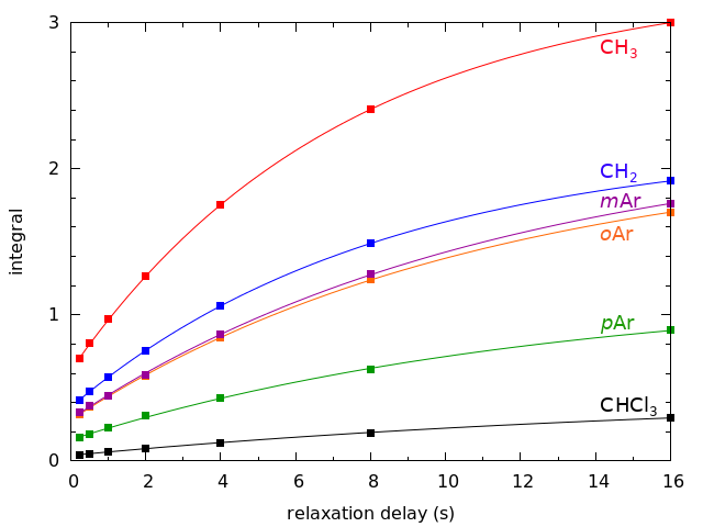
For the figure above the integral of the methyl resonance in the spectrum with the longest relaxation delay (16s) was set to three and used to calibrate all the other integrals. Looking at the curves its obvious that the integrals would increase further if the relaxation delay was increased. Slightly less obvious though, is that the integrals of the aromatic peaks are continuing to increase while those of the methylene and methyl are starting to flatten out. From the curves fitted to the integrals we can obtain rates for the curves. The rates for the two aliphatic peaks are similar, while the aromatic rates are all similar and slightly slower, and the chloroform rate is the slowest of all. These rates are a mixture of the T1 and T2 relaxation times of the signals and each signal has a different relaxation time that depends on its atom's magnetic environment.
For maximum intensity and the most accurate integrals it is generally recommended that the relaxation delay be set to five times the T1 relaxation time. The T1 relaxation time of the ethylbenzene methyl group has been reported as 17.4s1, requiring a relaxation delay of 87s. This is the fastest relaxing proton in ethylbenzene so theoretically the relaxation delay should be even longer to get accurate integrals of the other signals. A relaxation delay of more than 90s is not very practical, however. The usual solution to this problem is to reduce the relaxation delay and the pulse width. The shortened relaxation delay will reduce the accuracy of the integrals but still provide reasonable estimates. Reducing the pulse width will reduce the signal intensity, but will also reduce the time needed to return to equilibrium. The standard 1D 1H parameters at the SSPPS NMR Facility use a relaxation delay of 0.5s and a 30o pulse. For more accurate integrals the relaxation delay can be increased.
References
1. "NMR relaxation",
https://chem.ch.huji.ac.il/nmr/techniques/other/t1t2/t1t2.html
No comments:
Post a Comment